

Clinical Anatomy
Anatomy
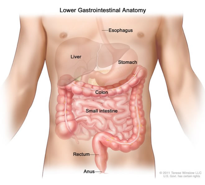
Colorectal surgeons treat diseases of the large intestine (bowel), comprising the colon, rectum and anus. The large intestine is part of the digestive system (gut), which is a series of hollow organs joined by a long inverted U-shaped muscular tube (over 10 metres long) starting at the mouth and finishing at the anus. The digestive system breaks down food and delivers nutrients to every cell in the body via the bloodstream. The organs comprising the digestive system are the stomach, small intestine, pancreas, liver and large intestine. The large intestine is made up of the colon and rectum (together about 2 metres long). These are kept in place in the abdomen by folds of soft tissue known as mesenteries, which are composed of fatty connective tissue containing blood vessels, nerves, lymph nodes and lymph vessels. The colon is attached to the back wall of the abdomen by a mesentery known as the mesocolon. The rectum is surrounded by a mesentery called the mesorectum.
Colon
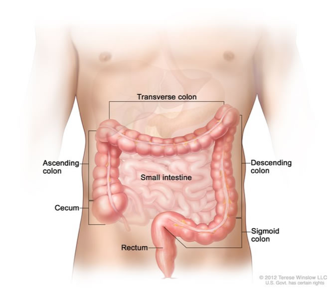
The colon receives partly digested food from the small intestine, absorbs water and salt, and propels waste (stool or faeces) to the rectum. It is the longest part of the large bowel, and has five parts:
- Caecum – this is the beginning of the colon and is located in the right lower abdomen.
- Ascending colon – this is found on the right side of the abdomen. It starts at the caecum and ascends (runs upwards) to a bend known as the hepatic flexure.
- Transverse colon – this comes after the ascending colon and hepatic flexure. Transverse means “across”. This part of the colon extends across the abdomen from right to left. At the end of the transverse colon is a bend known as the splenic flexure.
- Descending colon – this follows the transverse colon and splenic flexure, and descends (goes downwards) on the left hand side of the abdomen.
- Sigmoid colon – this is the final part of the colon, and is an S-shaped connection between the descending colon and the rectum. It is located on the bottom left hand side of the abdomen.
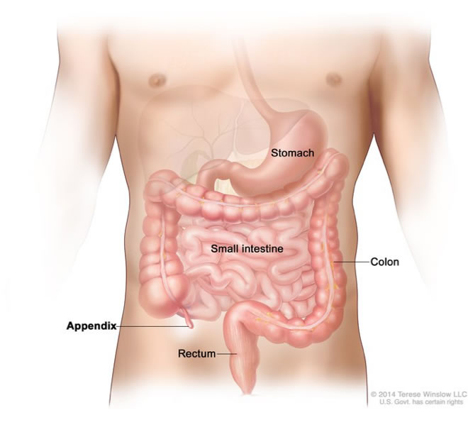
Appendix
The appendix is a small structure projecting from the caecum. In humans, the appendix has no known function and is thought to be left over from an earlier time in our evolution. If the appendix becomes infected or inflamed (a condition known as 'appendicitis'), it usually needs to be removed by surgery.
Rectum
The rectum is connected to the sigmoid colon and is the lowest part of the large intestine. It is about 15 cm long. The rectum receives stool (faeces) from the colon and stores it until it can be passed from the body via the anus.
Anus
The anus, or anal canal, is the opening at the bottom of the rectum through which faeces leaves the body, and is about 2–3 cm long. The anal canal and anal opening is surrounded by two muscular sphincters that regulate the movement of faeces. The internal sphincter is found just beneath the anal canal lining and is made up of circular layers of muscle that are not under voluntary control. Surrounding the internal sphincter is the external sphincter, which is under voluntary control. The external sphincter can be relaxed and contracted at will, except during the first few years of life when it is not yet fully developed. When stool arrives the bottom of the rectum and the top of the anus, nerve impulses transmitted from the anus cause involuntary relaxation of the internal sphincter. We then voluntarily contract the external sphincter until we are ready to go to the toilet. Once in the toilet, we relax the external sphincter muscle allowing the passage of stool. If going to the toilet is not convenient, the stool moves back up the rectum away from the anus. We lose the sensation of the need to pass stool and the internal muscle contracts again.
- Follow me on


